Introduction
In modern molecular biology, molecular imaging and gel documentation are two essential tools that allow scientists to visualize DNA, RNA, and proteins. These techniques help researchers understand biological processes, confirm experimental results, and share precise visual data. From PCR analysis to protein electrophoresis, these visualization methods have revolutionized how we study molecular structures.
What Is Molecular Imaging?
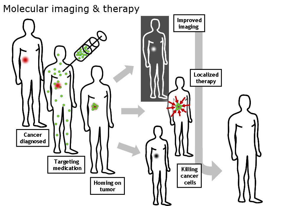
Molecular imaging is a group of techniques used to detect, track, and analyze molecules inside biological samples. It helps scientists visualize how genes, proteins, and metabolites behave within cells and tissues.

Key points:
It reveals molecular interactions, enzyme activity, and gene expression.
It is widely used in genetics, biotechnology, and cell biology.
Common imaging systems include fluorescence imaging, chemiluminescence, and bioluminescence.
With modern imaging systems, researchers can study complex molecular reactions in real time without harming the samples.
What Is Gel Documentation?
Gel documentation systems (also called gel imagers or gel doc systems) are used to record and analyze results from agarose or polyacrylamide gels after electrophoresis. These systems capture images of separated DNA bands, RNA fragments, or proteins, making it possible to measure their size and intensity. Read more
Core components:
UV or blue-light transilluminator: illuminates fluorescent dyes like ethidium bromide or SYBR Safe.
High-resolution camera: captures precise gel images.
Image analysis software: quantifies band intensity and molecular weight.
This technology ensures accurate visualization and digital storage of gel results for molecular biology workflows.
Applications in Molecular Biology
Molecular imaging and gel documentation play a vital role in a variety of laboratory applications:
PCR product visualization
DNA and RNA quantification
Protein separation and analysis (SDS-PAGE)
Cloning and plasmid verification
Gene expression studies
These systems enhance accuracy, improve reproducibility, and speed up data interpretation for researchers worldwide. Learn more
Advantages of Modern Gel Documentation Systems
High image sensitivity and resolution
Automatic exposure control for consistent results
Compatible with multiple fluorescent stains
Safe, digital, and easy to share or store results
Compared to traditional manual methods, modern gel documentation ensures cleaner, faster, and safer workflows in molecular labs.
The Future of Molecular Imaging
Recent advances combine AI-based image analysis and cloud data sharing for faster interpretation and collaboration. Portable imaging devices and software-driven automation are now bringing molecular visualization closer to real-time research and diagnostics.
Conclusion
Molecular imaging and gel documentation are indispensable tools in life science research. They bridge the gap between molecular data and visible results, allowing scientists to “see” biology in action. Whether you’re studying gene expression or verifying PCR results, these techniques make every discovery visible — one band at a time.